Our Services
As a fully licensed and equipped optometric practice, The Eye Site offers a complete range of eye care services to all our patients.
Whether the eye care issue involves correcting refractive errors with eyeglass or contact lenses, helping a student find amazing frames, or diagnosing/treating eye conditions and diseases, our experienced team will identify and implement the best eye care solutions for you.
Eye Health Evaluation
With our years of experience in diagnosing and treating typical vision disorders such as nearsightedness, farsightedness, amblyopia, presbyopia, cataracts, macular degeneration, and diabetic retinopathy, Dr. Ryan Nielson, Dr. Ana Nielson, Dr. Melissa Fowler, and Dr. Renee Rowley and their team are equipped to provide appropriate therapeutic medical eye care.
At the same time, the The Eye Site team offers a wide array of high-quality eye care products at reasonable prices. Our patients never pay too much for the best quality in eyeglasses, contact lenses, sunglasses, progressive and bifocal lenses, and outstanding service.
Treatment of Eye Disease
If you are diagnosed with an eye disease, you want the best treatment available to get your eyes healthy again. At The Eye Site, we stay current with best treatment practices. Based on your diagnosis, we may recommend a wide variety of approaches, including improved nutrition, prescription medicines, therapy and vision exercises, or medical procedures.
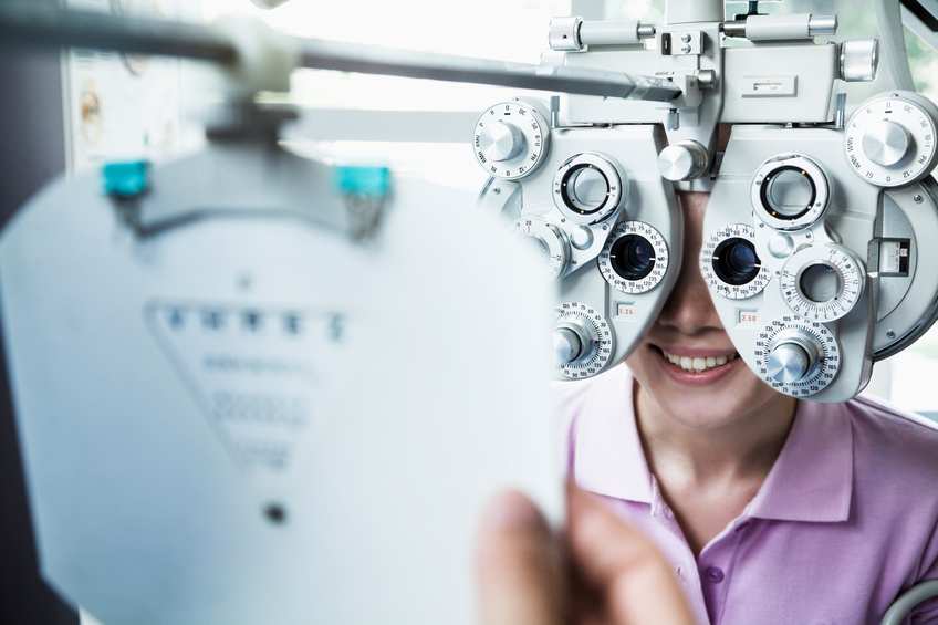
Good Eye Care Begins With A Yearly Eye Exam!
Although many do not realize it, the best way to protect your vision is with a yearly eye checkup. Even a basic eye exam can instantly detect many health-threatening conditions, such as tumors, vascular irregularities, and diabetes-related injury to the retina. At The Eye Site, that basic checkup is brief and painless.
Dry Eye Treatment
What is Dry Eye Syndrome?
Dry Eye Syndrome, also known as Ocular Surface Disease (OSD), is the most common eye disorder, affecting about 20% of the population. It is caused when one or all of the components of the tear film are not in balance. Clogged oil glands along both upper and lower lid margins contribute to 86% of ocular surface disease progression and dry eye. Poor lacrimal gland function further reduces the prodution of our "emergency eye wash tears." Damage to our goblet cells that reside in the tissue near our iris can reduce the output of our mucous tears which help nourish and protect our sight. Understanding the complex makeup of our tears is important in devising a proper treatment approach to solving OSD. We have cutting edge diagnostic and treatment technology to assess and manage every level of OSD.
What are the symptoms?
The most common symptoms of Dry Eye Syndrome include:
- Redness
- Irritation
- Scratchiness
- Burning
- Watery Eyes
Advanced dry eyes may damage the front surface of the eye and impair your vision. Depending on the severity of your symptoms, we have effective treatment solutions for OSD.
What are the risk factors?
Dry Eyes can develop for many of the following reasons:
- Age (a majority of people 40 and higher experience symptoms of dry eye)
- Gender (women are more likely to develop dry eyes due to hormonal changes)
- Medications (i.e., antihistamines, decongestants, blood pressure medications, and antidepressants can reduce tear production)
- Environmental conditions (i.e., screen time, exposure to smoke, wind, dry climates can increase tear evaporation and failure to blink can also contribute to dry eyes)
- Long-term use of contact lenses
- Refractive eye surgeries
How is Dry Eye Treated?
We start with a full examination and carefully examine the ocular surface with specific diagnostic equipment to determine which area of the tear film are in distress. Our doctors will then formulate an initial treatment plan which may include the following:
- Lid margin hygiene to encourage better oil gland production
- Discussion of supplements that may be beneficial
- Possible prescription eye drop medication
- TempSure Envi procedures to revitalize the oil glands along the lid margins
- ICON IPL (intense pulse light therapy) to reduce redness and vessel inflammation around the eyes and Demodex eradication
- Amniotic membrane technology
- Scleral lenses
- Autologous serum eye drops
- Or a combination of some of these strategies
Radio frequency treats dry eye by stimulating clogged tear glands.The oil producing tear glands reside along the upper and lower eyelids. These glands become blocked over time and lose their ability to produce the rich oil layer of the tear film. When this happens the eyes start to feel dry, irritated, burning and red. Over time these plugged glands can lead to atrophy (death of the glands), lid laxity and ocular surface disease and dry eye. As this condition worsens it can impact the ability to see clearly. This non-invasive treatment utilizes radio frequency technology to safely and effectively treat dry eyes and dermatochalasis (droopy eyelids) for a smoother, healthier-looking appearance. Best of all, there is no surgery or down time!
ICON IPL (intense pulses of light therapy) is an innovative treatment for ocular rosacea and dry eye. Initially found to be beneficial for treating facial rosacea for the past 20 years it has been more recently used in the dry eye field.
Countless studies show its effectiveness at reducing fine ocular rosacea along the upper and lower lid margins.Minimizing these pro-inflammatory vessels in the eyelids reduces the expression of toxic mediators that cause further destruction of the oil glands in the eyelids. Utilizing this technology is a game changer in treating ocular surface disease and dry eye.
Myopia Management
What is Myopia?
Myopia is a disease that affects the ability to clearly see objects far away. This is because the shape of the eyes causes the light rays to ben incorrectly, resulting in images being focused in front of the retina instead of on the retina. Myopia is progressive condition happens when the child is growing. If left untreated, the condition will worsen. Over the years, more and more children are diagnosed with myopia. While myopia cannot be cured, it can be prevented from worsening. Studies show that early intervention can slow or even stop the progression of myopia through a variety of proven treatments.
Here are the treatments that we offer:
Orthokeratology (Ortho K):
These overnight lenses are specifically designed to be worn only in the evening as your child goes to bed. They are taken out in the morning before your child prepares for the day. These lenses are custom made to comfortably reshape the cornea. Each child will undergo a consultation to receive their lenses. Studies have shown that myopia progression can be slowed and, in some cases, stopped through this treatment. Your child will be wearing these contacts at home. If your child wears the lenses every night, there would be no need for glasses or contacts during the day, as they can see clearly during the day.
Soft Mulitfocal Contacts:
By wearing the soft multifocal lenses, your child no longer has to depend on glasses. These contacts are meant to be worn during the day during school and in other various activities and should be taken out before bed time. What makes these lenses one-of-a-kind is their optical design, which is specifically crafted to provide clear vision during the day, yet slow down the progression of myopia at the same time.
Atropine Drops:
Our doctors may recommend a prescription eye drop to treat myopia. As everyone's eyes are different, they would recommend a specific time to apply these drops. While there will always be risks involved with any medical treatments, our doctors ensure these risks are minimized by having follow up appointments and monitoring the progress. Our doctors would usually recommend these drops for children who are showing rapid progression of myopia.
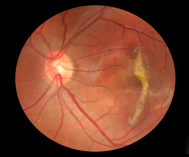
Optical Coherence Tomography / OCT
When patients come to our office for eye exams, many times there is testing that we do to help us diagnose problems. One of these tests uses Optical Coherence Tomography or OCT. Pictures taken with our OCT machine are generated by light waves that reflect off the back of the eye or retina, creating images similar to what could be produced by a low-power microscope. The OCT also provides cross-sectional images. These images can display the various layers of the retina. This technology is also used to image the optic nerve, which is important in glaucoma treatment and management.
OCT is a non-invasive and no-contact test that doesn't require preparation from the patient. There is no exposure to radiation since the machine uses light to obtain the images. The patient sits in front of a machine, a couple of bright flashes like a normal camera flash go off, and then the photos can be viewed on the machine within a minute.
The OCT is an extremely valuable tool used to help diagnose and manage common retinal eye diseases such as macular degeneration, macular edema (fluid in the retina), and macular hole/epiretinal membranes. We use the initial OCT images to aid in making a definitive diagnosis. The OCT compares these initial images of your eye to a database of images of normal eyes matched to your age. In this way, the first OCT images we take can help point out potential issues if there is something that looks different from normal or average. Subsequent OCT images can then compare how you look now compared to how you looked initially. This can be very valuable in gauging how well treatment is working or if the problem is progressing.
The other common use of the OCT is for monitoring and managing glaucoma. We usually take initial images of the optic nerve, and the OCT can then compare these images to those of age-matched healthy control patients. The OCT will usually be repeated every year so we can follow any changes or progression over time.
The advent of OCT has revolutionized the way we evaluate the retina because we can now detect subtle findings not otherwise easily seen during clinical exams. This makes the OCT one of the most valuable tests we can do in our office.
LASIK
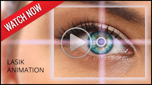
LASIK, a form of refractive surgery, is an popular option for vision correction, often eliminating the need to wear glasses or contact lenses. Simply put, LASIK reshapes the cornea with a laser.
Other surgical alternatives have become available. Among these is a technique called phakic IOL implantation which involves implanting a lens behind the cornea, but in front of the iris. With this new option, many of those who were too highly nearsighted for LASIK are now candidates for refractive surgery.
If you are interested in refractive surgery, please let us know. Refractive surgery is not to be taken lightly. Detailed testing is necessary to determine whether or not you are a good candidate for the surgery. If testing shows you to be a good candidate, we can help you choose the refractive surgeon who is most appropriate for your case. In addition, we provide post-operative care for refractive surgery.
Corneal Refractive Therapy (CRT)
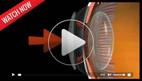
Corneal Refractive Therapy (CRT) is a great advancement in refractive therapy options. CRT allows you to see clearly during the day without glasses or contact lenses. Specially designed therapeutic lenses gently reshape the front surface of your eye while you sleep.
Imagine great vision all day without contacts, glasses, or surgery. No more problems from dust, allergies, or dryness. CRT has also been shown to slow the progression of nearsightedness in children and teenagers.
Previously, if you were nearsighted and wanted to see clearly during the day, your options were glasses, contacts, or laser surgery. CRT offers a non-surgical solution. No more worry about broken frames or torn contact lenses. Additionally, while LASIK is usually recommended only for those over 18 years of age, CRT is ideal for our younger patients who are active in sports and do not like the hassle of glasses or contacts.
For more information on Corneal Refractive Therapy, call today to schedule your FREE CRT consultation or visit Paragon at www.paragoncrt.com.
Optomap Retinal Exam
In our continued efforts to bring the most advanced technology available to our patients, Dr. Ryan Nielson, Dr. Ana Nielson, Dr. Melissa Fowler, and Dr. Renee Rowley are proud to announce the inclusion of the Optomap Retinal Exam as an integral part of your eye exam.
Many eye problems can develop without warning and progress with no symptoms. Early on, you might not notice any change in your vision. However, diseases such as macular degeneration, glaucoma, retinal tears or detachments, as well as other health problems such as diabetes and high blood pressure, can often be detected with a thorough exam of the retina. The retina is the part of your eye that catches the image of what you are looking at, similar to the film in a camera.
An Optomap Retinal Exam provides:
- A scan to confirm a healthy eye or detect the presence of disease.
- An overview or map of the retina, giving your eye doctor a more detailed view than he can achieve by other means.
- The opportunity for you to view and discuss the Optomap images of your eye with your doctor at the time of your exam.
- A permanent record for your medical file, enabling your optometrist to make important comparisons if potential problems show themselves at a future examination.